Diagnosis: intussusception
This patient presents with a history typical intussusception: pain that is colicky, severe, and intermittent. Other typical symptoms include currant jelly stools (bloody mucous) and lethargy between pain paroxysms. What is not typical about this patient is his age, as only 5% of intussusception cases occur in adults.1
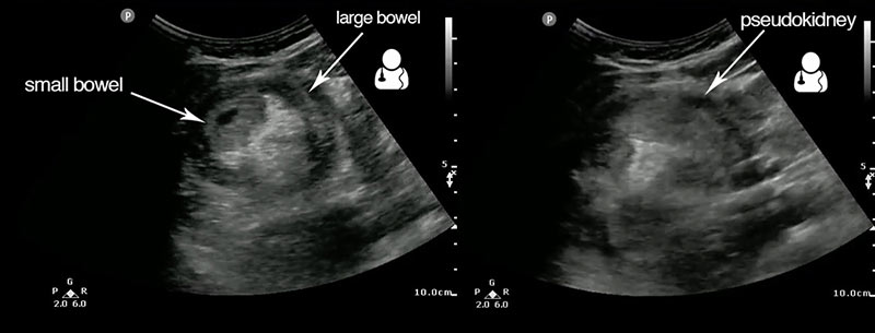
This abdominal ultrasound demonstrates the two classic findings for intussusception: the target sign appearance of bowel in the transverse plane, and the “pseudokidney” appearance of the bowel in the oblique plane.2 This extensive ileocecal intussusception resulted in the target sign’s visibility from the right lower to right upper quadrant of the patient’s abdomen.

Take home points:
- Provider-performed bedside ultrasound has been shown to be 90% sensitive for intussusception in the pediatric setting.3
- Another study found that junior radiology resident-performed scans were as sensitive as radiology attending scans at identifying intussusception.4
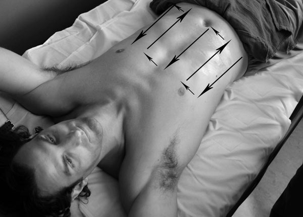
- When evaluating the abdomen for bowel pathology, the provider should employ a systematic scanning technique to avoid missing pathology. One such method is described as “mowing the lawn.5“

1. Marinis A, Yiallourou A, Samanides L, et al. Intussusception of the bowel in adults: a review. World J Gastroenterol. 2009;15(4):407-11. [PDF]
2. Gingrich AS, Saul T, Lewiss RE. Point-of-care ultrasound in a resource-limited setting: diagnosing intussusception. J Emerg Med. 2013;45(3):e67-70. [article]
3. Eshed I, Gorenstein A, Serour F, Witzling M. Intussusception in children: can we rely on screening sonography performed by junior residents?. Pediatr Radiol. 2004;34(2):134-7. [article]
4. Chang YJ, Hsia SH, Chao HC. Emergency medicine physicians performed ultrasound for pediatric intussusceptions. Biomed J. 2013;36(4):175-8. [PDF]
5. Dawson, Mallin. Introduction to Bedside Ultrasound, Volume 2. 2013. Apple iBook. [iBook]


