80 yo f presents from home with complaints of pain around her g-tube. The g-tube has been functioning properly, and the patient denies pain with feeding. Her past medical history is significant for diabetes, peripheral vascular disease and 2 MI’s in the past. She denies fever/chills, diarrhea. Physical exam shows normal vitals signs. Inspection of area around g-tube shows mild redness extending approximately 1-2 cm around the g-tube. Movement of the g-tube exposes 2-3 maggots surrounding the area.
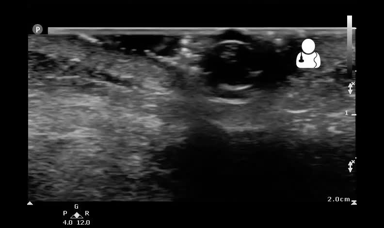
An ultrasound machine was used to identify the extent of the maggot infestation, and the following images were obtained.

The clips recorded demonstrated a much more extensive infestation of the maggots, and showed the maggots to only be present along the tract of the g-tube and did not appear to extend past into the peritoneum, or past the margin of the g-tube tract.
- Myiasis = maggot infestation
- There are many different types of myiasis, including wound myiasis, subcutaneous myiasis, gastrointestinal myiasis, ophthalmic myiasis, aural myiasis, body orifice associated myiasis.1
- Wound myiasis appears to be the most common, and the most common species identified is the green botfly.1
- Factors associated with myiasis include homelessness, peripheral vascular diasease, coronary artery disease, stroke, those with circulatory compromise, alcoholics and diabetics.1 Immunocompromised states appear to be unrelated to myriasis.1
- Treatment includes thorough cleaning of the area, proper wound care, tetanus prophylaxis, and antibiotics if bacterial infection is suspected.1
- Previous case reports of ultrasound being used to aid in the diagnosis of myiasis have been reported, but these described subcutaneous myiasis.2,3 To the knowledge of this author, no previous reports have been published describing the use of ultrasound to aid in the work-up of a patient with wound myiasis.
Management of this patient included a surgery consult. They scrubbed the area with Dakin solution until there were no more maggots visible. The patient was instructed to keep the area clean and dry and to clean it once a day with the Dakin solution.
Scan and Post by: Jacob Avila, MD, RDMS
- Sherman RA. Wound myiasis in urban and suburban United States. Arch Intern Med. 2000;160(13):2004-14. [PDF]
- Schechter E, Lazar J, Nix ME, Mallon WK, Moore CL. Identification of subcutaneous myiasis using bedside emergency physician performed ultrasound. J Emerg Med. 2011;40(1):e1-3. [pubmed]
- Bowry R, Cottingham RL. Use of ultrasound to aid management of late presentation of Dermatobia hominis larva infestation. J Accid Emerg Med. 1997;14(3):177-8. [PDF]


