A 19-year-old female presents with right hip pain that was first noticed after a ground-level mechanical fall. PMHx significant for IVDU. Her vital signs are as follows: T°98F HR 105 (sinus) BP 130/80 RR 18. On examination she has pain with any motion of the right hip. XR does not demonstrate any evidence of fractures or effusions.
Bedside ultrasound of the affected hip is performed and demonstrates the following:
What is the diagnosis?

Septic Hip Effusion
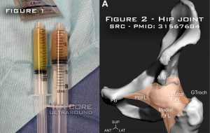
This ultrasound demonstrates a hip effusion on the affected side. On further questioning, the patient did describe a soreness to the right groin that had been worsening over the course of a few weeks. After successful ultrasound-guided aspiration of the effusion (See Figure 1), the patient was admitted for IV antibiotics and orthopedic consultation.
 The hip joint capsule attaches to the pelvis proximally, envelops the femoral head and neck, and attaches distally to the intertrochanteric line1. (SeeFigure2:)
- The ultrasound transducer should be placed along the axis of the femoral neck, with the probe marker facing in a cephalo-medial direction.
- A small amount of fluid can be normally seen in the hip joint capsule. If the total size is >7mm or or a difference between the affected and non-affected side of >1 mm, it is considered abnormally enlarged2.
For a tutorial on how to perform the examination, check out the 5 minute sono video on the hip exam
References
- Ng KCG, Jeffers JRT, Beaulé PE. Hip Joint Capsular Anatomy, Mechanics, and Surgical Management. The Journal of bone and joint surgery. American volume. 2019; [PMID: 31567684]
- Koski JM, Anttila PJ, Isomäki HA. Ultrasonography of the adult hip joint. Scandinavian journal of rheumatology. 1989; 18(2):113-7. [PMID: 2660254]


