43 y/o female complains of 3 days of paroxysmal right upper quadrant abdominal pain associated with nausea and vomiting.

Diagnosis: Normal Gallbladder
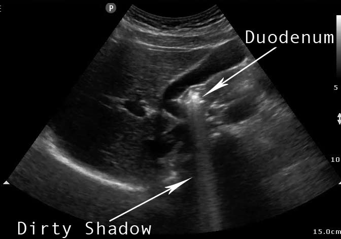
Although this patient did present with symptoms suggestive of biliary colic, this ultrasound demonstrates a normal thin-walled gallbladder. It can be noted that there is a hyperechoic extralumenal structure deforming the gallbladder posteriorly with some mixed hyper/hypoechoic shadowing. This most likely represents a loop of small bowel (probably duodenum); there is no evidence of gallstones.
Take home points:
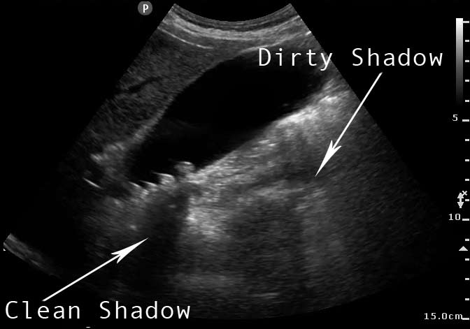
- Gallstones are typically seen as dependent, echogenic structures with a deeply hypoechoic (black) shadow.
- Bowel loops commonly masquerade as stones, but typically have a “dirty shadow” appearance.
- Two simple ways to differentiate loops of bowel from stones are 1) watch carefully for peristalsis and 2) reposition the patient in left lateral decubitus – usually the loop of bowel will be pulled away giving you a cleaner view of the patient’s gallbladder.
- Be careful not to mistake the duodenum for a wall echo shadow sign (WES). If you suspect a WES sign, reposition the patient as above and rescan.


