This patient presents with skin redness and warmth on his right lateral thigh for 3 days. On exam there is a 4 cm patch of erythema, with a moderate amount of induration. Should this be incised?

Answer: Abscess, Incise and Drain
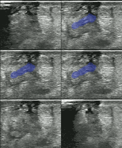
This scan, obtained with a high frequency linear transducer demonstrates increased subcutaneous fluid,  classically described as cobblestoning. At first glance, there is no clear hypo-echoic fluid collection. However, when compression is applied with the probe there is a swirl of iso-echoic purulent material evident in the subcutaneous tissue.
- When presented with the clinical question of abscess vs cellulitis, ultrasound has been shown to change the gestalt management frequently,1,2,3 about half of the time in one study.4
- Usual scan technique includes using a high frequency transducer in a high resolution preset (superficial or MSK), fanning through the affected area.
- In pure cellulitis, early findings show an increased thickening and echogenicity of subcutaneous tissue. As cellulitis progresses, you see the classic cobblestoning appearance (although this finding is not specific to cellulitis and can be seen with general cutaneous edema).5
- A hypoechoic fluid collection is typically diagnostic of abscess, but sometimes a lymph node can be interpreted as positive. A lymph node will have a slightly hyperechoic center and generally will demonstrate color flow when doppler is applied.6
 In this case, no hypo-echoic fluid collection was demonstrated. Compression revealed the answer. To improve the clinician’s sensitivity for subcutaneous fluid collections, a series of gentle compressions should be performed every 1-2 cm throughout the cellulitis, in a fashion similar to DVT evaluation..
- To the left is a second example of significant iso-echoic fluid collection made obvious by compression.
 If you like the ultrasound education you get from UOTW, you will absolutely love CastleFest, a world class ultrasound event held in April. Whether you’re an ultrasound novice or want to hone your experienced skills, come eat, drink and learn with the best educators in the field: Dawson, Mallin, Weingart, Mallemat, and more. Oh, and I’ll be there too. Register now.
- Iverson K, Haritos D, Thomas R, Kannikeswaran N. The effect of bedside ultrasound on diagnosis and management of soft tissue infections in a pediatric ED. The American journal of emergency medicine. 30(8):1347-51. 2012. [pubmed]
- Marin JR, Bilker W, Lautenbach E, Alpern ER. Reliability of clinical examinations for pediatric skin and soft-tissue infections. Pediatrics. 126(5):925-30. 2010. [pubmed]
- Giovanni JE, Dowd MD, Kennedy C, Michael JG. Interexaminer agreement in physical examination for children with suspected soft tissue abscesses. Pediatric emergency care. 27(6):475-8. 2011. [pubmed]
- Tayal VS, Hasan N, Norton HJ, Tomaszewski CA. The effect of soft-tissue ultrasound on the management of cellulitis in the emergency department. Academic Emergency Medicine. 13(4):384-8. 2006. [pubmed]
- Chao HC, Lin SJ, Huang YC, Lin TY. Sonographic evaluation of cellulitis in children. Journal of ultrasound in medicine : official journal of the American Institute of Ultrasound in Medicine. 19(11):743-9. 2000. [pubmed]
- Dawson M, Mallin M, Introduction to bedside ultrasound volumes 1 + 2. [iTunes]


