Answer: Posterior tibial nerve block
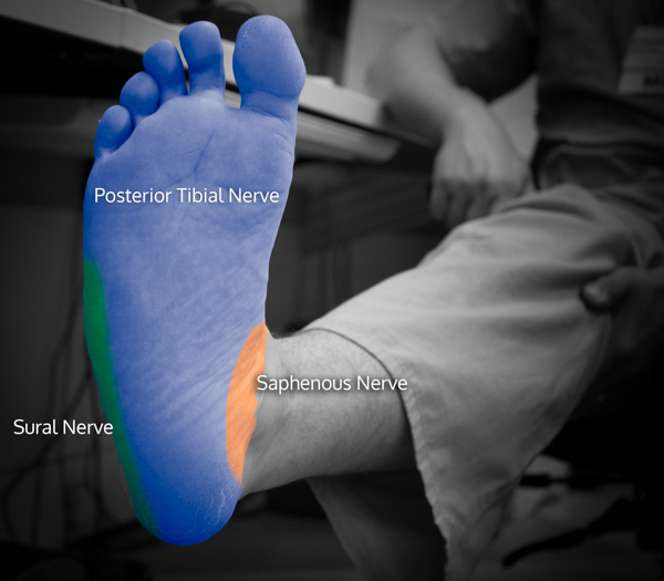
- There are 5 nerves to the foot below the ankle, which include posterior tibial, sural, deep and superficial peroneal nerves (which are from the sciatic nerve) and a branch of the femoral nerve which is from the saphenous.
- The sole of the foot is predominantly innervated by the posterior tibial nerve.

- There are two methods that can be used for effective sensory blockade of the sole of the foot, and they include landmark based (LM) and US based (US).1
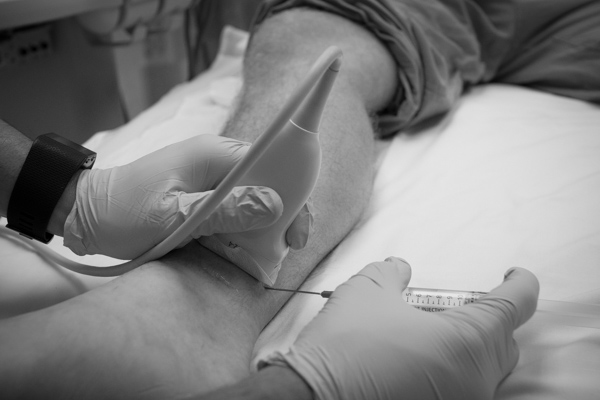
- In the LM approach, a needle is inserted posteriorly to the posterior tibial artery just above the medial malleolus. 1
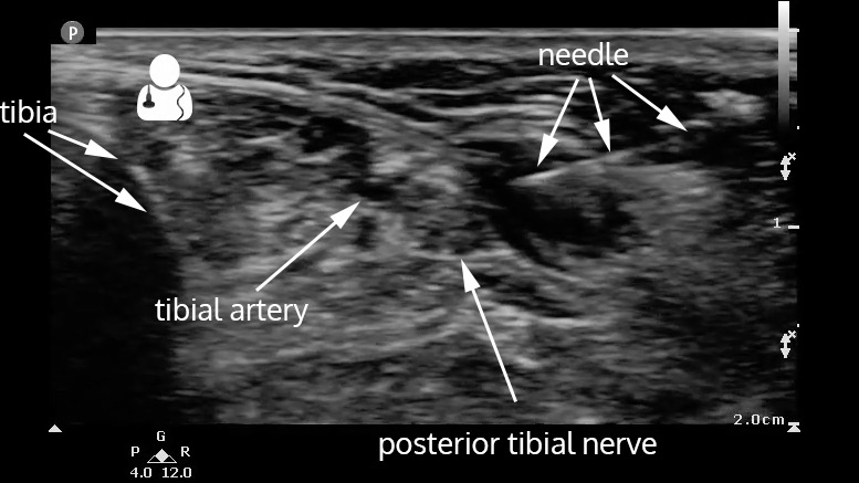
- For the US approach, a linear transducer should be placed in a similar location to the LM approach, identifying the posterior tibial artery and the posterior tibial nerve.


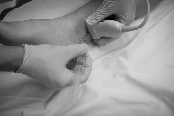
- Both an in-plane and out-of-plane technique can be used.


- If an in-plane technique is used, the patient can be placed in the prone position to facilitate needle advancement. The needle should be placed just anterior to the achilles tendon.

- Approximately 5 mL of local anesthetic should be visualized to surround the nerve, taking special care to not inject any inside any blood vessels.
- Instead of aiming your needle for the nerve itself, aim for an adjacent spot within the fascial plane where the nerve runs. As anesthetic is injected, the nerve can be seen separating away from the nerve sheath and facial plane. You can then advance your needle above and below the nerve to surround it. Inject, advance, inject, advance…
- Here is an example of an in plane approach, used on our patient here:
- Redborg KE, Antonakakis JG, Beach ML, Chinn CD, Sites BD. Ultrasound improves the success rate of a tibial nerve block at the ankle. Reg Anesth Pain Med. 2009;34(3):256-60. [pubmed]


