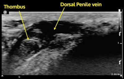
Diagnosis: Mondor’s Disease
Bedside ultrasound visualized a non-mobile hyperechoic clot in the proximal end of the dorsal penile vein.
- Mondor’s disease is defined as a thrombosis of a superficial vein, classically located on breast tissue.1
- Superficial thrombosis of the dorsal penile vein was first described in 1958.2
- Literature on this disease consists of cases and case series, as it is a fairly rare disease. It has been associated with local trauma, abstinence, urogenical infections and clotting disorders, among others.1
- The diagnosis is typically made on physical exam, but ultrasound can be used to aid in the diagnosis.3
- The ultrasound transducer of choice is the linear transducer. The dorsal penile vein should be evaluated along its entirety. The visualization of a hyperechoic cloth within the lumen can help confirm the diagnosis. Generally, b-mode visualization is all that is needed, but assistance with color Doppler may be necessary.
- Conservative medical treatment, including the use of NSAIDs and warm compress has been found to be efficacious, but thrombectomy or superficial penile vein resection may be indicated if refractory.4,5
- Girardi L. Mondor’s disease affecting the superficial dorsal vein of the penis. VASA. 2012;41:(3)233-5. [pubmed]
- Helm JD, Hodge IG. Thrombophlebitis of a dorsal vein of the penis; report of a case treated by phenylbutazone (butazolidin). J Urol. 1958;79:(2)306-7. [pubmed]
- Hamilton J, Mossanen M, Strote J. Mondor’s Disease of the Penis. West J Emerg Med. 2013;14:(2)180. [pubmed]
- Al-Mwalad M, Loertzer H, Wicht A, Fornara P. Subcutaneous penile vein thrombosis (Penile Mondor’s Disease): pathogenesis, diagnosis, and therapy. Urology. 2006;67:(3)586-8. [pubmed]
- Kumar B, Narang T, Radotra BD, Gupta S. Mondor’s disease of penis: a forgotten disease. Sex Transm Infect. 2005;81:(6)480-2. [pubmed]


