The patient is a 65 y/o female who presents from home with severe shortness of breath, weakness and confusion per EMS. Family is enroute, EMS has no further history at this time. Vitals: 78/30, 140, 28, 82% RA, 99.6F. What is the diagnosis and treatment?

Answer: Cardiogenic shock, norepinephrine
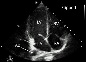
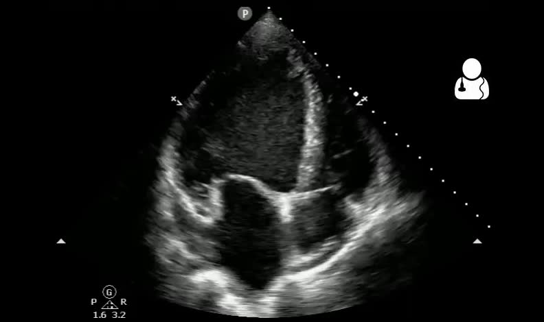
Respect the probe marker. This bedside echocardiogram is inadvertently reversed. Fortunately for this patient, the error was recognized and a correctly-oriented apical view was obtained:
There is profound global left ventricular systolic dysfunction noted, with bowing of the interventricular septum into the right ventricle. The left atrium appears dilated as well. The right ventricle is diminutive compared to the left ventricle.
- When performing a bedside echocardiogram for undifferentiated shock, it is imperative that you have the probe marker oriented correctly. You may be inclined to diagnose this patient with signs of acute pulmonary hypertension, massive pulmonary embolism and give thrombolytics unnecessarily.
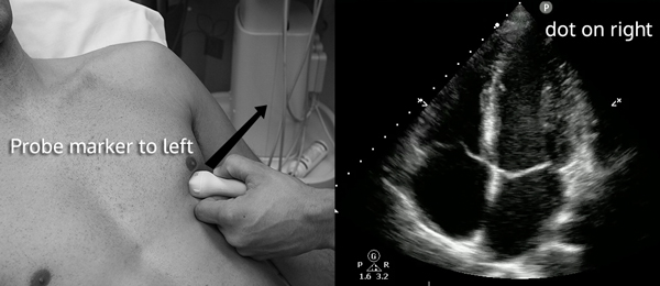
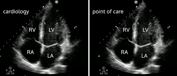
- When it comes to probe marker and dot orientation for an apical 4 chamber view, there is often a discrepancy between cardiology and point-of-care echocardiography. If the dot positioned on the left side of the screen (typical of point-of-care scans), the probe marker should point to the patient’s right:

- However, if the dot is positioned on the right side of the screen (cardiology standard), the probe marker should be directed toward the patient’s left:

- Please do note that the resulting image is exactly the same:

- There is no reason to memorize/learn both of these orientations. You should just remember one set of echocardiogram probe/dot orientations and do it that way all the time. Most ultrasound machines can be set to place the dot on either side of the screen for cardiac mode, just make sure you keep it consistent.

- When you orient the probe incorrectly, there are a couple clues to help you realize this error. First, if you lay the probe down on the chest from your apical view, you will get an apical five chamber view. The aortic outflow tract will be located off the left ventricle.

- A second way to tell if you have your apical view orientation incorrect is to find the descending aorta. This landmark corresponds to the left side of the heart.
- Lastly, the tricuspid valve tends to be slightly more apical than the mitral valve, but this difference may be subtle (2mm on average).1
- Otto CM. Textbook of Clinical Echocardiography, Expert Consult – Online and Print. Elsevier Health Sciences; 2013.


